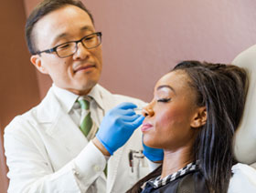
Depending on the severity and the type of skin cancer you have, we will recommend one or more of a wide range of treatment options. We may also use these treatments if you have Actinic Keratosis. Actinic keratoses (AKs), are precancerous lesions that typically develop on fair-skinned individuals in areas exposed to sunlight.They are characterized by rough, scaly patches that may resemble stubborn scabs that resist shedding and can even bleed when irritated. Roughly 10% to 30% of these lesions can evolve into malignancies if left untreated.
Preventing actinic keratosis and skin cancer hinges on effective sun protection strategies, such as using a broad-spectrum sunscreen with SPF 30 or higher daily, wearing a wide-brimmed hat to protect vulnerable areas like the ears, and wearing long sleeves or UPF-rated clothing.
Excision (Malignancies on bodily areas)
Mohs micrographic surgery is a highly specialized treatment used for skin cancers found on the head, neck, or cosmetically sensitive areas. However, the mainstay of treatment for most skin cancers is surgical excision.The cancerous tumor is excised with some of the surrounding healthy tissue and is then submitted to our laboratory for an evaluation of margins. Excision has a high cure rate and can reduce the risk of the cancer recurring.
Electrodessication & Curettage (Superficial skin cancers)
This treatment provides great results and is less invasive than many other skin cancer procedures. A curette is employed to scrape away the cancerous tissue until healthy tissue is revealed. Next, an electric current is applied to the area, effectively destroying any remaining cancer cells and controlling bleeding through cauterization of the wound. The residual effect from electrodessication and curettage is typically a hypopigmented or light-colored scar usually around the size of a quarter depending on the size of the initial skin cancer.
Prescription Medication (Superficial BCCs or AKs)
Chemotherapy for the skin works to destroy mutated cells present in actinic keratoses or superficial basal cell carcinoma (BCC) cancers. Based on your symptoms, needs, and budget, we will work with you to find the best prescription medication for you. Two of the most common prescription medicines we use are Imiquimod and 5-Fluorouracil. Imiquimod works by stimulating the body’s immune response to target and eradicate abnormal skin cells. 5-Fluorouracil interferes with cancer cells’ ability to replicate DNA, ultimately leading to their death and the regression of skin cancer lesions. Both of these creams are used for about two to three weeks at a time. Common side effects include redness, irritation and sometimes blistering. These side effects can be calmed down with the use of a low-potency topical steroid.
Cryotherapy (AKs)
Cryotherapy involves the application of extreme cold, typically using liquid nitrogen, to freeze and destroy abnormal cells in the skin affected by actinic keratosis. During the procedure, the liquid nitrogen is sprayed directly onto the lesions, causing them to blister and eventually slough off as the treated skin heals.
Chemical Peels (AKs, Sun damage)
Moderate chemical peels, such as the Vitalize Peel or Perfect Peel, can be utilized to exfoliate away actinic keratoses. These work by burning off the damaged cells to improve the quality and health of the skin. We will work with you to find the best chemical peel based on your needs, symptoms, and budget.
Laser Treatment
Laser skin resurfacing is another treatment for moderate-to-severe actinic keratosis. The top layer of skin (epidermis) is ablated or removed. Heat from the laser penetrates into the second layer of skin (dermis) to stimulate an intense collagen reaction. The combination of ablating the epidermis and stimulating deep collagen production removes actinic keratoses and results in healthy, rejuvenated skin.
Superficial Radiation Therapy (SRT)
Superficial Radiation Therapy (SRT) is a non-invasive treatment option that delivers low-energy radiation directly to the skin’s surface, targeting cancerous cells while preserving surrounding healthy tissue. Patients receive short, 30-second treatments once or twice a week over several weeks.
Photodynamic Therapy
Ameluz is a topical gel used to treat actinic keratosis (AK) and sun-damaged areas on the face and scalp. The gel is applied to lesions and allowed to absorb for a period of time. Next, a special deep-penetrating red light is used to activate the medication. Most patients will see their lesions disappear over the next several weeks.